
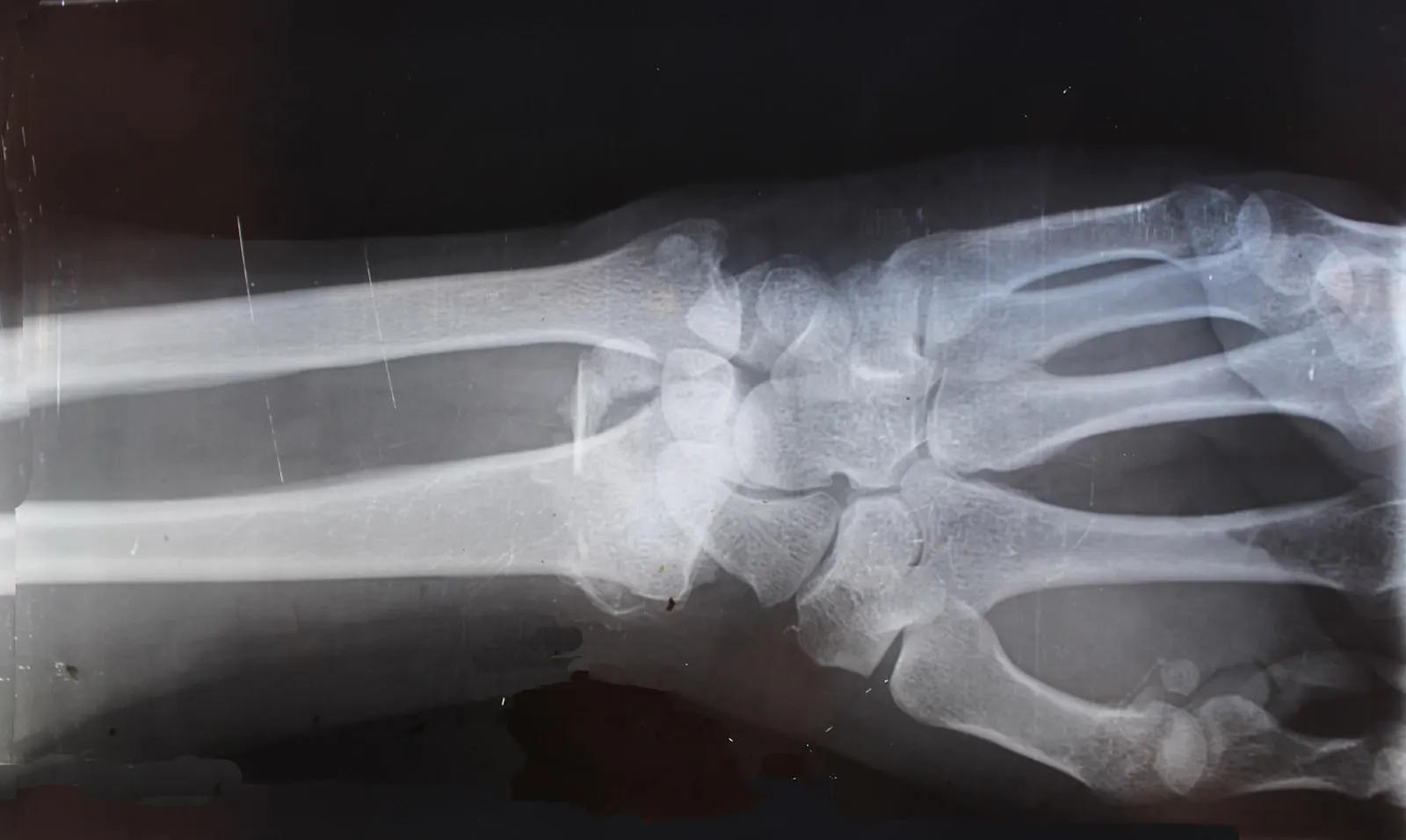
An exam reaches as far as hands and ears can go. Imaging shows the rest. The exact position of a fracture, a swallowed toy, a mass on the liver or the litter growing in a mother's belly can only be judged clearly with X-ray and ultrasound.
When is an X-ray needed?
For bone and joint problems, X-ray is where we turn first. We use it to locate fractures and dislocations after trauma such as traffic accidents and falls, to evaluate the joints of a limping patient, to search for swallowed foreign objects and to examine the chest cavity. On our digital system the image appears on screen within seconds, with no film to develop.
When is an ultrasound needed?
Ultrasound is the gold standard for soft tissue, meaning the internal organs. The liver, kidneys, bladder, spleen and intestines are assessed in real time. Pregnancy monitoring is one of its happiest uses. We watch the little heartbeats together.
Ultrasound is painless and completely harmless. Most patients lie calmly through the whole procedure and no anesthesia is needed. At most, the fur over the area being examined may need a trim.
An image alone is not enough
Imaging earns its value through sound interpretation. We weigh the images against the exam and laboratory findings, and as we explain, we show you on the screen exactly what we are seeing. Seeing it with your own eyes makes every step of the process easier to follow.